
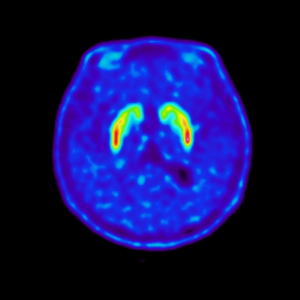
Somatotopy in Parkinson’s disease
"Somatotopy" refers to how areas of the brain are organized according to the body part they affect. The striatum is the brain region that coordinates complex thinking and movement. Plasticity refers to changes in connections within the brain, which can happen to make up for changes that are related to PD. This study aims to assess these changes in connections within the brain in Parkinson's disease (PD) using PET (Positron Emission Tomography) and fMRI (functional Magnetic Resonance Imaging) on the Hybrid PET/MRI scanner. PET and fMRI imaging together allow investigate changes in the striatum in people living with Parkinson's disease when compared to people without Parkinson’s disease.
Recently published: https://www.centreforbrainhealth.ca/news/new-study-reveals-how-the-brain-reorganizes-itself-in-early-parkinsons-disease/
Principal Investigator: Dr. A. Jon Stoessl
Contact: stoessl.research@ubc.ca
-
Title: Functional segregation in Parkinson’s disease
Authors: Dongning Su et al.
Background and Objectives
It is widely known that Parkinson’s disease affects dopamine, an important chemical messenger involved in movement, thinking, motivation, and reward. To understand how dopamine is affected, it is helpful to first understand the brain structures involved. Parkinson’s disease affects a group of interconnected structures deep within the brain called the basal ganglia. These structures help us select, start, and control movements, but are also involved in other functions such as learning, motivation, decision-making, and reward.
A main part of the basal ganglia is the striatum, which includes two important regions called the caudate and putamen. The striatum receives signals from many areas of the brain and is particularly influenced by dopamine. Dopamine helps regulate activity within these brain circuits and allows different parts of the basal ganglia to work together.
Many of the dopamine-producing cells that supply the striatum are located in a region of the brain called the substantia nigra. These cells send dopamine to the striatum, helping regulate the activity of the basal ganglia. In Parkinson’s disease, many of the dopamine-producing cells are lost. As a result, there is less dopamine signaling in the striatum, particularly areas involved in movement.
However, dopamine release is organized differently across different parts of the striatum. Different parts of the basal ganglia are connected with different brain networks and contribute to different functions. This raises an important question: Does Parkinson’s disease only change how much dopamine is available, or does it also change how dopamine release is organized across different parts of the striatum?
Key Findings
Different movements produced different dopamine patterns
In healthy participants, different movements produced different patterns of dopamine release across the striatum when compared to people with Parkinson’s disease. For example, finger tapping and foot tapping produced distinct patterns associated with the upper and lower limbs. This organization of different areas according to different parts of the body is called “somatotopy.” The researchers also examined “functional segregation”, which describes how different functions are associated with distinct patterns of activity in different areas of the brain. In this study, movement, thinking, and reward produced different patterns of dopamine release across the striatum in healthy participants.
Parkinson’s disease changes the organization of dopamine release
In people with Parkinson’s disease, these patterns were less clearly separated and more overlapping. Instead of different activities producing distinct patterns of dopamine release in particular areas, dopamine release was more widespread across regions of the striatum. This means that Parkinson’s disease may affect not only how much dopamine is available, but also where and how dopamine release is organized. It was also found that the clear separation between upper- and lower-limb movements seen in healthy participants was not present in the same way in people with Parkinson’s disease.
Some brain areas may help compensate for dopamine loss
Interestingly, some areas of the striatum that are less affected by Parkinson’s disease showed increased dopamine release during movement. The researchers suggest that these areas may be helping to compensate for dopamine loss in other parts of the brain.
Changes in dopamine organization extend beyond movement
The changes were not limited to movement. The researchers also looked at dopamine release while participants completed the card-sorting and reward tasks. In healthy participants, these different activities produced their own segregated patterns of dopamine release. In Parkinson’s disease, these patterns were less clearly separated and showed greater overlap. This suggests that changes in the organization of dopamine release may involve not only movement-related circuits, but also circuits involved in thinking, decision-making, and reward.
Broader Implications
The findings from this study provide a new way of thinking about how Parkinson’s disease affects the dopamine system.
1. Parkinson’s Disease May Be More Than Dopamine Loss
Parkinson’s disease is often described as a condition in which the brain loses the ability to produce dopamine. This study suggests that the story may be more complicated. The brain may not only produce less dopamine, but the dopamine that remains may also be released in different patterns across the striatum depending on what a person is doing.
2. Different Brain Functions May Become Less Separated
In a healthy brain, different movements and activities appear to produce relatively distinct patterns of dopamine release. In Parkinson’s disease, these patterns become more overlapping. The researchers suggest that this reduced separation may make it more difficult for the brain to select the specific movement or behaviour that is needed at a particular time. This may contribute to some of the difficulties people with Parkinson’s disease experience when switching between movements or managing more than one task at a time.
3. The Brain May Try to Adapt
The increased dopamine release seen in some less-affected areas of the striatum may represent an attempt by the brain to adapt to dopamine loss. This suggests that some changes seen in Parkinson’s disease may not necessarily be harmful. Some may represent the brain trying to maintain function despite the effects of the disease.
4. Understanding These Changes May Help Future Research
Understanding how dopamine release changes across different areas of the striatum may provide researchers with a better picture of why Parkinson’s disease affects people in different ways. Rather than looking only at how much dopamine has been lost, future research may also benefit from understanding how the remaining dopamine system is organized and how the brain adapts to dopamine loss.
Conclusion
This study adds to our understanding of Parkinson’s disease by showing that the effects of the disease may involve more than simply losing dopamine.
In healthy participants, different movements, thinking tasks, and reward experiences produced relatively distinct patterns of dopamine release across the striatum. In people with Parkinson’s disease, these patterns became more widespread and overlapping, meaning that the different activities were no longer represented by such clearly separated patterns.
These findings suggest that Parkinson’s disease may affect both dopamine release and the way dopamine release is organized throughout the striatum. An important takeaway is that increased dopamine release in some less-affected areas may represent the brain attempting to compensate for dopamine loss elsewhere.
Together, these findings contribute to a more complete picture of how the dopamine system changes in Parkinson’s disease and may help guide future research into the difficulties with movement, thinking, and managing multiple activities that can occur with the condition.
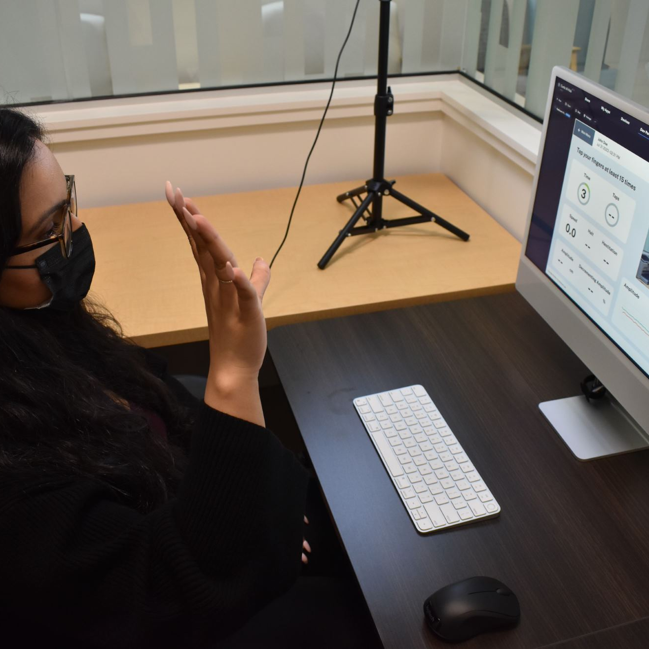
Confidential Automatic Monitoring, Examination, and Recognition of Disease Activity (CAMERA) study
Patients with neurodegenerative disorders such as Parkinson’s disease (PD) often make long journeys to visit their physicians and trial multiple therapies and medications to manage their symptoms. Physicians use clinical assessments to inform drug regimens for their patients, however these assessments are performed infrequently, and symptoms may change vastly in between visits. This research aims to create a video-based data collection platform specifically designed to assess PD symptoms by capturing detailed facial expressions, hand movements, finger tapping, and eye movements. Using advanced machine learning algorithms, the collected data is analyzed to generate comprehensive, data-driven reports for clinicians and researchers. This will enhance the understanding of PD symptoms and offer an efficient, scalable method for assessing disease severity through non-invasive video-based assessments.
Principal Investigator: Dr. Martin McKeown
Contact: mckeown.lab@ubc.ca
-
Title: Impact of Video Quality on Video-based Digital Assessment of Parkinson’s Disease
Authors: Atefeh Irani, Kye Won Park, Maryam S. Mirian, Mahdi Shakouri, Michael Grundy, Juana Ayala, Reshad Hosseini, Hadi Moradi, Martin J. McKeown
Summary
Remote video analysis is increasingly used to measure Parkinson’s disease motor symptoms at home. These recordings allow clinicians and researchers to track symptoms such as slowed or reduced movement more frequently, and in some cases from a patient’s own home. This approach, often called using “digital biomarkers,” has the potential to make monitoring more convenient and more reflective of everyday life. However, home recordings are not always captured under ideal conditions, and differences in lighting, camera setup, or internet limitations may affect video quality.
In this study, our researchers examined how video quality influences the accuracy of automated Parkinson’s assessments. We analyzed finger-tapping videos collected both in clinic settings and in real-world home environments, and systematically tested how factors such as video resolution, frame rate, lighting, and camera positioning affected the system’s ability to track hand movements and estimate clinical scores.
We found that two factors were especially important for reliable results: recording speed and clear visibility of the hand. Videos recorded at fewer than 24 frames per second significantly reduced measurement accuracy, and recordings where the hand appeared too small or too large within the frame also increased errors. Encouragingly, extremely high video resolution was not necessary, even lower-resolution videos produced reliable results when recording speed and positioning were appropriate.
These findings help establish simple, practical guidelines for patients recording videos at home, supporting accurate and clinically meaningful remote assessments. By identifying the key requirements for reliable recordings, this work helps advance safe and scalable video-based monitoring of Parkinson’s disease, bringing symptom tracking closer to patients’ everyday environments while maintaining clinical quality.
-
Title: Facial expression analysis to uncover the relationship between sialorrhea and hypomimia in Parkinson’s disease
Authors: Eline Serbée, Kye Won Park, Atefeh Irani, Maryam S. Mirian, Juana Ayala Castaneda, Michael Grundy, Martin J. McKeown
Link: https://www.frontiersin.org/journals/neurology/articles/10.3389/fneur.2025.1661043/full
Summary:
Drooling (medically called sialorrhea) and reduced facial expression (hypomimia) are two features commonly seen in Parkinson’s disease (PD). While both can affect quality of life, they are often under-recognized in clinic. This study looked at how these two symptoms relate to each other using both traditional clinical scales and automated video-based facial analysis.
Researchers recorded short videos of facial expressions from 52 people with PD and used artificial intelligence to measure specific features of facial movement around the mouth and eyes. They also used a validated questionnaire to assess how much drooling individuals experienced. Clinical rating scales showed a modest link between reduced facial expression and more severe drooling. When facial video features were analyzed with computer algorithms, the relationship was stronger with certain patterns in facial movement correlating moderately with drooling severity.
These findings suggest that automated facial analysis can help quantify the relationship between facial expressivity and drooling in PD. Because facial videos are easy to record, this approach could improve detection of non-motor symptoms that are otherwise hidden or under-reported. Ultimately, better recognition of these symptoms may lead to more comprehensive symptom monitoring and targeted care strategies for individuals living with Parkinson’s disease.
-
Title: HiLWS: A Human-in-the-Loop Weak Supervision Framework for Curating Clinical and Home Video Data for Neurological Assessment
Authors: Atefeh Irani, Maryam S. Mirian, Alex Lassooij, Reshad Hosseini, Hadi Moradi, Martin J. McKeown
Summary:
Video recordings are increasingly being used to help monitor movement symptoms in people living with Parkinson’s disease, allowing clinicians to assess changes more frequently and sometimes from patients’ homes. However, videos recorded outside the clinic often vary in quality due to differences in lighting, camera setup, or how tasks are performed, which can reduce the accuracy of automated assessments. In this study, researchers developed a new framework called HiLWS (Human-in-the-Loop Weak Supervision) that combines artificial intelligence with expert clinical input to improve how medical videos are prepared, labeled, and analyzed for remote neurological assessment.
Using more than 2,000 videos collected in both clinical and home environments, the team showed that real-world recording conditions significantly influence measurement accuracy. They identified practical guidelines for reliable recordings, including maintaining adequate recording speed and ensuring the hand is clearly visible in the frame, while demonstrating that very high video resolution is not required. The framework also improved consistency in symptom scoring by combining input from multiple clinical experts with machine learning models and directing expert review toward uncertain cases.
These findings help make remote Parkinson’s assessments more reliable and scalable, supporting more frequent monitoring outside the clinic while maintaining clinical quality. More broadly, the approach provides a foundation for building trustworthy AI tools for remote healthcare, helping expand access to neurological care and enabling continuous monitoring for people living with chronic conditions.
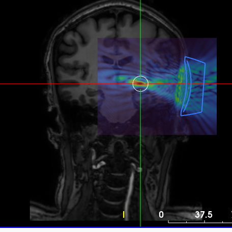
Low Intensity Focused Ultrasound for Parkinson’s Disease Tremor
In this study, we aim to compare the effects of targeting two different brain regions, the traditionally treated VIM and the zona incerta (ZI), using LIFUS for tremor control in Parkinson’s disease. Past research has shown that the ZI may be an important area for treating tremor and other Parkinson’s symptoms like stiffness and uncontrolled movements. Our goal is to understand how these brain regions contribute to Parkinson’s tremor and how the network responds to LIFUS. The knowledge gained will contribute to developing more effective treatments for Parkinson’s disease in the future.
Principal Investigator: Dr. Martin McKeown
Contact: mckeown.lab@ubc.ca
-
Title: Low-Intensity Transcranial Ultrasound Effects on the Ventral Intermediate Nucleus and Zona Incerta for Parkinson’s tremor
Authors: Maggie Q. Vuong, Varsha Sreenivasan, Soojin Lee, Ben Quinn, Martin S. Keung, Michael Grundy, Juana Ayala Castañeda, Hongchae Baek, Martin J. McKeown
Link: https://www.sciencedirect.com/science/article/pii/S1935861X26000021
Summary:
Tremor is a common and challenging symptom of Parkinson’s disease (PD) that does not always respond well to medication. While high-intensity focused ultrasound is an FDA-approved method used to reduce tremor by targeting a small brain region called the ventral intermediate nucleus (VIM), Parkinson’s tremor is complex because it can occur both at rest and while holding a posture. Another nearby brain region, the zona incerta (ZI), is also linked to tremor and connected to broader movement networks. In this study, we used low-intensity focused ultrasound (LIFUS), a non-invasive and temporary form of brain stimulation, to better understand how modulating brain activity in these two regions affects tremor in people living with PD.
Nineteen participants with PD took part in the study. Each person first underwent MRI scanning to precisely guide ultrasound targeting. On the same day, participants received two separate LIFUS sessions: one targeting the VIM and one targeting the ZI. Before and after each session, participants completed tasks designed to bring out tremor while researchers measured movement using small motion sensors placed on the arm. Functional MRI scans were also collected to examine how brain activity changed following each type of stimulation.
The results showed differences between the two brain targets. Stimulation of the VIM reduced tremor mainly when participants were holding a posture, which is consistent with previous research. In contrast, stimulation of the ZI mildly improved both postural tremor and tremor at rest, suggesting it may influence a wider network involved in movement control. Brain imaging supported this finding, showing broader changes in brain activity after ZI stimulation compared with VIM stimulation. Together, these findings suggest that while the VIM remains an effective target for certain types of tremor, the ZI may represent a promising new target for treating Parkinson’s tremor more broadly in the future.
Tau and Neuroinflammation Imaging in Parkinson’s Disease and Related Disorders
Parkinson's disease (PD) results from the loss of dopamine producing neurons in the brain; however, the mechanisms for the initiation and progression of this loss are still largely unknown. Recent research has indicated that tau may be a contributor to the development and progression of the disease. Using Positron Emission Tomography (PET), an in-vivo brain imaging technique, we hope to determine the presence and extent of tau accumulation in patients with Parkinson’s disease, Progressive Supranuclear Palsy, Multiple System Atrophy and Corticobasal degeneration.
Principal Investigator: Dr. A. Jon Stoessl
Contact: stoessl.research@ubc.ca
-
Title: Misfolded protein deposits in Parkinson's disease and Parkinson's disease-related cognitive impairment, a [11C]PBB3 study
Authors: Michele Matarazzo, Alexandra Pérez-Soriano, Nasim Vafai, Elham Shahinfard, Kevin Ju-Chieh Cheng, Jessamyn McKenzie, Nicole Neilson, Qing Miao, Paul Schaffer, Hitoshi Shinotoh, Jeffrey H Kordower, Vesna Sossi, A Jon Stoessl
Link: https://www.nature.com/articles/s41531-024-00708-z
Key Findings
Misfolded Protein Aggregation in PD
One of the study’s significant findings is the higher [11C]PBB3 binding in an area of the brain called the posterior putamen of PD subjects compared to healthy controls. This indicates an accumulation of misfolded proteins in this region, highlighting it as a critical site of protein aggregation in Parkinson’s disease. However, there was no significant difference found in [11C]PBB3 binding between PD subjects and healthy controls in another region, the substantia nigra, which is traditionally considered a primary site of dopaminergic neuron loss in PD.
Cognitive Impairment and Anterior Cingulate
The study revealed that CI-PD subjects show increased [11C]PBB3 binding compared to CN-PD and HC in another area of the brain, the anterior cingulate cortex. This higher binding indicates that greater misfolded protein deposition in this region is associated with more severe cognitive impairment. The results suggest that pathology in the anterior cingulate could serve as a significant marker for cognitive decline in PD, offering potential diagnostic and therapeutic targets for managing cognitive impairments in PD.
Neuroinflammation
The study also examined the relationship between misfolded protein deposits and neuroinflammation by comparing [11C]PBB3 and [11C]PBR28 binding in nigrostriatal regions. The results show no significant correlation between these two measures, suggesting that the neuroinflammation detected by [11C]PBR28 does not directly relate to the accumulation of misfolded proteins measured by [11C]PBB3. This finding highlights the complexity of PD pathology, where misfolded protein accumulation and neuroinflammation may contribute to the disease through distinct and perhaps independent mechanisms.
Broader Implications
The findings of this study have several important implications for understanding and treating Parkinson’s disease and its cognitive impairments:
Role of Striatal Axonal Terminals
The preferential involvement of striatal axonal terminals, particularly in the posterior putamen, in misfolded protein aggregation emphasizes the need to consider these regions in PD research and treatment strategies. This challenges the traditional focus solely on the substantia nigra and underscores the complexity of PD pathology.
Cognitive Decline and Anterior Cingulate Pathology
Identifying the anterior cingulate cortex as a critical area for cognitive impairment in PD provides a potential biomarker for diagnosing cognitive decline. This could lead to earlier and more accurate identification of patients at risk for cognitive impairment, allowing for earlier interventions and better management of cognitive symptoms.
Therapeutic Targets:
The study's insights into the specific regions involved in misfolded protein aggregation open new avenues for therapeutic development. Targeting the posterior putamen and anterior cingulate cortex with treatments aimed at reducing misfolded protein deposits could help slow disease progression and mitigate cognitive decline.
Diagnostic Imaging
The use of [11C]PBB3 PET imaging as a tool to visualize and quantify misfolded protein deposits represents a significant advancement in PD research. This imaging technique could be used to monitor disease progression, evaluate the efficacy of new treatments, and enhance our understanding of the underlying mechanisms of PD and its cognitive impairments.
Conclusion
The study adds to our understanding of how protein clumps affect PD and its cognitive effects. By identifying the posterior putamen as a major site of clumped proteins and showing the link between the anterior cingulate cortex and cognitive decline, the study highlights how different brain regions are involved in PD. It also shows that inflammation and protein clumps might not always go hand in hand, revealing the complexity of PD. PET imaging with tracers like [11C]PBB3 are invaluable for studying these issues, opening the door to better diagnostics and treatments in the future.
Precision Stimulation Project
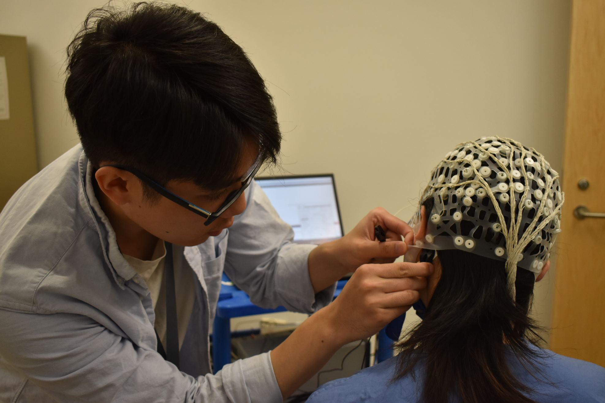
The Precision Stimulation study uses galvanic vestibular stimulation (GVS), a non-invasive stimulation technique that sends small electrical currents to selectively target the vestibular system. The vestibular system provides a sense of balance and information about one’s body position. While prior work has demonstrated improvements in both motor and non-motor symptoms of Parkinson disease with GVS, McKeown’s team believes that by customizing the stimulation parameters to the individual, the therapy has the potential to be much more effective.
This study combines GVS with electroencephalogram (EEG) and functional magnetic resonance imaging (fMRI) recordings. The information from the EEG and fMRI are then used to custom-design GVS stimulation parameters for that individual. The team is then seeing if such a customized stimulus is significantly better than a “one-size-fits-all” stimulus during the performance of motor tasks.
Principal Investigator: Dr. Martin McKeown
Contact: mckeown.lab@ubc.ca
-
Title: Individualised, brain-informed motor vigour assessment for targeted non-invasive brain stimulation in Parkinson’s disease
Authors: Varsha Sreenivasan, Martin S. Keung, Soojin Lee, Hanaa Diab, Wyatt Vercher, Martin J. McKeown
Summary
A common characteristic of slowness of movement (bradykinesia) in Parkinson’s disease (PD) is reduced motor vigour — the intensity, speed, and motivational drive that propel our movements. Current treatments, including deep brain stimulation and medications such as levodopa, primarily aim to improve bradykinesia. However, these treatments are usually evaluated using simple behavioural measures like reaction time or movement speed. These single measures vary markedly across individuals, and using one uniform measure across individuals is not optimal. Motor vigour is associated with the subjective motivational drive to move and is therefore an individual-specific attribute. In this work, we introduce an individualised, brain-based way of measuring motor vigour and test whether it can perform better than simple behavioural measures in detecting the effects of brain-modulating treatments in Parkinson’s disease.
We studied 18 people with Parkinson’s disease who performed a rapid, forceful hand-squeezing task. During the task, participants received electrical vestibular stimulation. Nine types of stimulation were tested, including a sham (placebo-like) condition. At the same time, brain activity was recorded using functional MRI. Participants performed this task once each before and after taking levodopa medication. We measured six aspects of behaviour, including reaction time, how long the squeeze lasted, and how quickly it was performed. We then analysed patterns of brain activity to identify three brain networks and examined how their responses changed over time. By combining brain activity and behavioural data for each individual using advanced statistical modelling, we created a personalised measure called individualised, brain-informed motor vigour (IndMV).
We show that traditional behavioural measures did not show a clear benefit of electrical stimulation compared to sham. Moreover, these measures could not distinguish between medication states very well (60–82%). On the other hand, the personalised IndMV measure outperformed the traditional behavioural measures by capturing significantly greater changes caused by stimulation and differentiating between medication conditions with 93% accuracy. When stimulation was optimised for each individual, motor vigour improved by more than 30%. IndMV is a new, personalised way of measuring motor vigour that goes beyond standard behavioural tests. By integrating brain and behavioural data, it more effectively captures the effects of brain modulation and offers a promising tool for better identifying and tailoring treatment effects in Parkinson’s disease.
-
Title: Rapid Profiling of EEG Responses to Non-Invasive Brain Stimulation in Parkinson’s Disease: A Biomarker-Driven Screening Framework
Authors: Sepideh Hajipour Sardouie, Mahdi Babaei, Mahsa Naseri, Servin Mehrtash, Mohammad Hossein Faramarzi, Zahra Kavian, Martin Keung, Varsha Sreenivisan, Hanaa Diab, Maryam S. Mirian and Martin J. McKeown
Link: https://www.mdpi.com/2227-9059/14/2/352
Summary:
Parkinson’s disease affects not only movement but also the brain’s natural rhythms. These rhythms can be measured using electroencephalography (EEG), a non‑invasive method that records electrical activity from the scalp. In recent years, researchers have become increasingly interested in whether gentle forms of brain stimulation can shift these rhythms towards a healthier pattern. However, finding the right stimulation settings for each person remains a major challenge.
In this study, we developed a fast and systematic way to test many different types of Galvanic Vestibular Stimulation (GVS) signals, over 300 in total, while recording EEG in people with Parkinson’s disease and in healthy volunteers. GVS uses very small electrical currents applied behind the ears to subtly activate the balance system, which in turn influences brain activity.
To understand how each stimulation pattern affected the brain, we created a combined “biomarker score” that captured several important features of EEG activity, including brainwave power, interactions between different brainwaves, and signal complexity. Instead of relying on movement or behavioral changes, this score allowed us to directly measure how close the brain’s response moved toward a healthier profile.
We found that several types of stimulation, particularly specific rhythmic patterns, consistently shifted EEG activity in a positive direction. Importantly, these effects occurred only during stimulation, with no lingering changes afterward. Although this was a pilot, exploratory study, the approach shows promise as a rapid screening tool for identifying personalized stimulation strategies. By focusing on brain biomarkers rather than symptoms alone, this framework may help guide the development of more targeted, individualized brain-modulating therapies for Parkinson’s disease.
-
Title: Comprehensive Benchmarking of EEG Biomarkers for Parkinson’s Disease: Robustness, Reliability, and Clinical Discriminability Across Multiple Datasets
Authors: Sepideh Hajipour Sardouie, Mahsa Naseri, Mahdi Babaei, Servin Mehrtash, Mohammad Hossein Faramarzi, Zahra Kavian, Maryam S. Mirian and Martin J. McKeown
Summary:
Parkinson’s disease affects millions of people worldwide, yet diagnosing and tracking its progression remains challenging. One promising tool is electroencephalography (EEG), a simple, non‑invasive method that measures the brain’s electrical activity. Many research groups have proposed EEG‑based “biomarkers” for Parkinson’s, but their findings often disagree. This inconsistency makes it difficult to know which brain signals truly reflect the disease and which are too sensitive to differences in data processing or analysis.
Our study takes an important step toward solving this problem. We performed the first large, systematic comparison of 16 commonly used EEG biomarkers across two independent datasets of people with Parkinson’s and healthy adults. We tested how stable each biomarker is when researchers change key analysis choices, such as how much denoising is applied, how long the data segments are, or how signals from multiple electrodes are combined. We also examined how reliably these biomarkers can distinguish Parkinson’s patients from healthy individuals, and how well they capture changes when patients are on or off their medication.
We found that some biomarkers, especially those describing the brain’s background activity (aperiodic features), as well as measures of signal complexity and variability, were consistently robust and clinically informative. In contrast, biomarkers based on rhythmic brain activity were much more sensitive to methodological choices and less reliable in recordings of the brain at rest. By identifying which EEG features are truly dependable, our work provides practical guidance for researchers and clinicians. This benchmarking framework can help standardize EEG analysis and accelerate the development of reliable brain‑based markers for diagnosing Parkinson’s, monitoring symptoms, and evaluating new treatments.
Exercise and Brain Energetics in Parkinson’s Disease
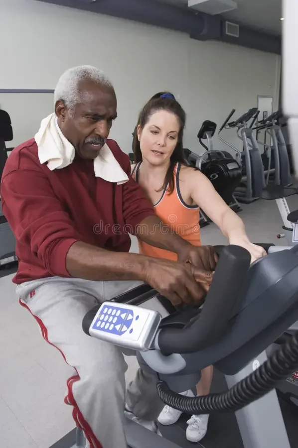
It is well known that exercise helps slow down disease progression, but how exactly does it work? What changes occur in the brain in response to sustained levels of exercise? This research study aims to answer these questions using positron emission tomography (PET) and magnetic resonance imaging (MRI) to study brain energetics, or how the brain produces and uses energy. There is evidence that PD alters healthy brain energetics, and exercise may be a means to slow down or even reverse these changes.
We are currently recruiting individuals participating in less than 120 minutes of high-intensity exercise per week. You will be asked to come in for assessments, an exercise test, and a single PET/MRI scans, and then repeat the three visits after 6 months. You may be reimbursed for travel or provided with transportation.
Click here for more information
Principal Investigator: Dr. Vesna Sossi
Contact: jess.mckenzie@ubc.ca
Parkinson’s Disease Biomarker Study
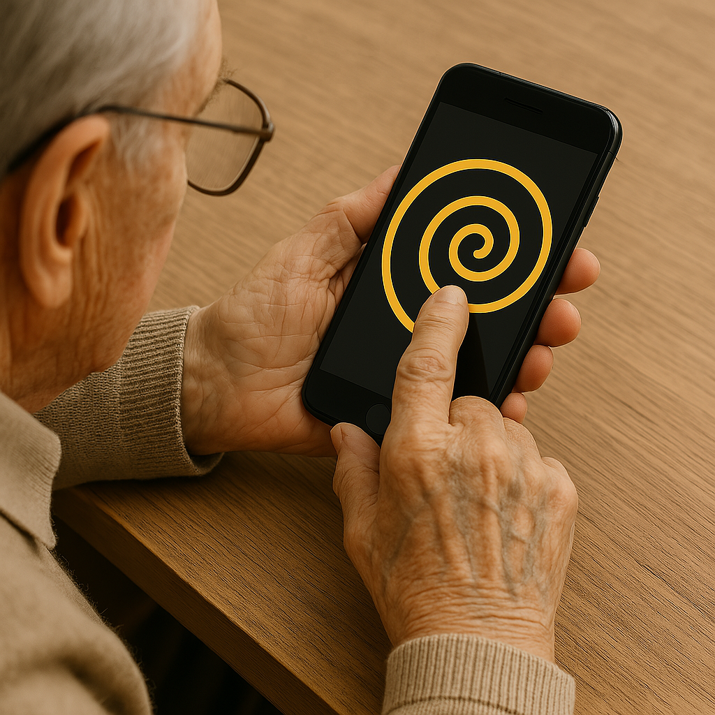
Monitoring characteristic motor changes in individuals diagnosed with Parkinson’s Disease (PD) and individuals at risk of developing PD (prodromal PD) remains a challenge as motor symptoms fluctuate subtly every day. Moreover, the instances that patients are able to see their doctors is limited, contributing to these difficulties in monitoring. This study aims to determine if the Roche PD Mobile Application can be used to reliably and remotely monitor motor and non-motor symptoms in PD through active tests and passive monitoring. Active tests are designed to assess symptoms using short daily tasks on a smartphone, while passive monitoring assesses mobility throughout the day.
Analysis in progress
Principal Investigator: Dr. Martin McKeown
Contact: mckeown.lab@ubc.ca

Music PD
Listening to different types of audio, such as music or podcasts, have been shown to improve brain health outcomes in healthy and clinical populations; these effects, however, have yet to be tested in Parkinson’s disease. This study aims to examine the effects of audio interventions, such as music and podcasts, on targeting apathy (lack of motivation for daily tasks) in people with Parkinson’s disease. Participants will take part in an 8-week intervention period involving either music or podcast listening, and 3 in-person visits involving clinical assessments and functional/structural MR scans in the MRI scanner, for a total of 12 weeks of involvement.
Analysis in progress
Principal Investigator: Dr. Silke Appel-Cresswell
Contact: miguel.jose@ubc.ca

GBA Multimodal Study
Mutations in the GBA gene could be, in some cases, associated with Parkinson’s disease. Understanding the mechanisms that lead to the development of the disease in people who have the GBA mutation may have implications for identifying therapeutic targets in both genetic and non-genetic cases. In this Michael J. Fox Foundation sponsored study, participants with this mutations in this gene will be asked to complete several PET/MR scans and assessments of movement, mood, and thinking.
Analysis in progress
Principal Investigator: Dr. A. Jon Stoessl
Contact: stoessl.research@ubc.ca
Individualized prediction of medication wearing off using wearables
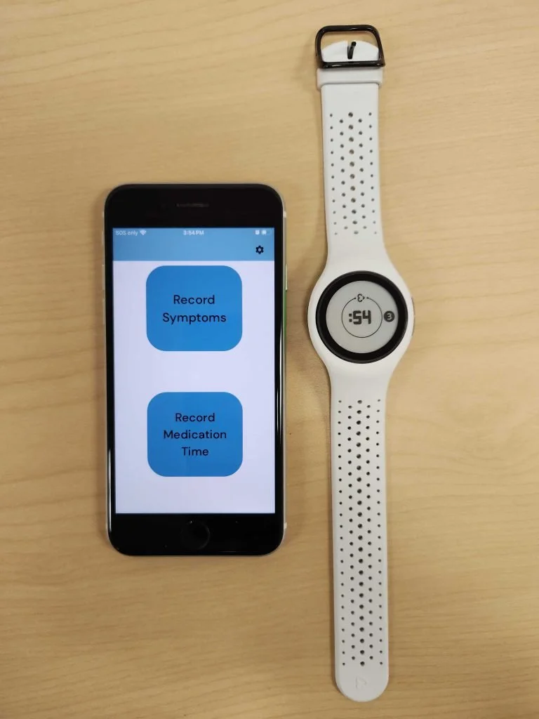
Parkinson’s disease is most commonly treated with levodopa, and while this treatment method has shown to significantly help reduce symptoms, many patients also experience “wearing-off” (WO) episodes, where symptoms re-emerge prior to their next dose. This study aims to investigate whether these episodes can be predicted – and ultimately prevented – using machine learning to assess biomarkers from the autonomic nervous system such as electrodermal activity (EDA), a measure of sweating from the skin. To monitor EDA and WO episodes, participants will be asked to wear a wrist sensor for 72 hours and complete questionnaires on an hourly basis via their mobile device.
Analysis in progress
Principal Investigator: Dr. Martin McKeown
Contact: mckeown.lab@ubc.ca

Impact 360 for Parkinson’s Disease
This study looks to examine the neuroprotective effects of exercise, nutrition, and mindfulness within older healthy individuals and those diagnosed with Parkinson's disease. Participants will be screened and, if successful, enrolled in a 6-month intervention consisting of exercise, nutrition and mindfulness classes. Participation in the study will average approximately five hours per week. Study visits include blood work, DEXA scan, MRI, and questionnaires.
Analysis in progress
Principal Investigator: Dr. Silke Appel-Cresswell
Contact: impact.360@ubc.ca
Recent Publications
Functional segregation in Parkinson’s disease. Su, D., Hanania, J.U., Stoessl, A.J.S., et al. (2026). Science Advances. doi:10.1126/sciadv.aed170
Anxiety is associated with increased risk of suicidality in Parkinson’s disease. Lam, J.S., Tosefsky, K.N., Zhu, J., et al. (2026). Journal of Parkinson’s Disease. 2026;0(0). doi:10.1177/1877718X251410887
A randomized safety and feasibility crossover trial of two Mediterranean-ketogenic interventions in individuals with Parkinson’s disease. Tosefsky, K, Lam, J.S., Wang, Y.N., et al. (2026). Journal of Parkinson’s Disease. 2026;0(0). doi:10.1177/1877718X261418986
Individualising galvanic vestibular stimulation further improves visuomotor performance in Parkinson’s disease. Menon, A., Vigneswaran, M., Zhang, T., Sreenivasan, V., Kim, C., & McKeown, M. J. (2025). Bioengineering, 12(5), 523. https://doi.org/10.3390/bioengineering12050523
EEG dynamical features during variable-intensity cycling exercise in Parkinson’s disease. Alizadeh, Z., Arasteh, E., Mirian, M. S., Sacheli, M. A., Murray, D., Appel-Cresswell, S., & McKeown, M. J. (2025). Frontiers in Human Neuroscience, 19, 1571106. https://doi.org/10.3389/fnhum.2025.1571106
Disease-Modifying Trials in Treated Parkinson’s Disease: “Stable Treated” Does Not Equate with Biological Stability. Mouradian, M. M., Stoessl, A. J., & Lang, A. E. (2025). Movement Disorders, 40(9), 1778–1790. https://doi.org/10.1002/mds.30259. PMC 12485585
Sex and gender differences in the molecular etiology of Parkinson’s disease: considerations for study design and data analysis. Schaffner, S. L., Tosefsky, K. N., Inskter, A. M., Appel-Cresswell, S., & Schulze-Hentrich, J. M. (2025). Biology of Sex Differences, 16(1), 7. https://doi.org/10.1186/s13293-025-00692-w
A generalized framework for in vivo detection of dopamine release using positron emission tomography. Hanania, J. U., Bevington, C. W. J., Cheng, J. K., et al. (2025). Journal of Cerebral Blood Flow & Metabolism. Published online 19 Sept (2025). https://doi.org/10.1177/0271678X251362958